Categorias
Posterior Capsular Rupture Management

PCR management – Right and Wrong management

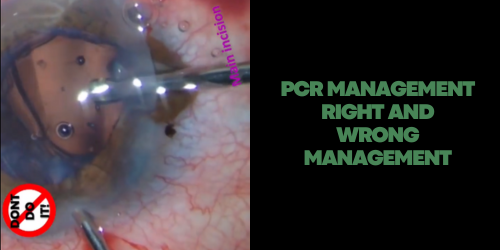
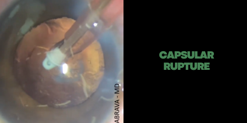
In PCR we recommend pars plana vitrectomy or to create New Small Incision in the Cornea. In this management is essential:
– Don’t use the main incision.
– The vitrectomy through the main incision tends to lose more vitreous. – The use of triamcinolone to better vitreous visualization, making the anterior vitrectomy more effective.
– A scleral incision should be created 3.0 – 3.5 mm posterior to the limbus to access the vitreous behind in pars plana vitrectomy.
– The vitreophagus tip is used in maximum cutter.
– There are two modes to set the vitrectomy. The “Cutting – I/A” and the “I /A – Cutting”. The first one is indicated for the vitrectomy, because the machine “cut” the vitreous first. The second one is indicated after the vitrectomy has been performed. The rule is to set to the highest speed cutter, however, the aspiration, intraocular pressure, and vacuum must be set to lower limits. – Once the vitreous has been removed from the anterior chamber and the incisions, the next step is to switch to the I/A Cutting mode to remove the remaining cortex.
– The vitrectomy through the main incision tends to lose more vitreous – Parameters: 23G tip: Bottle height: 75cm / Vacuum: 150-200mmHg / Flow rate: 10-15cc/min / Cutting: maximum.

Categorias
Posterior Capsular Rupture Management

Capsular Rupture

It is a patient with posterior polar cataract. During the plaque removing… Capsule Rupture! What to do?

1. OVD to avoid vitreous loss;
2. 26g needle to create new pivot;
3. Posterior rhexis;
4. OVD again;
5. IOL with captured optic zone.

Categorias
Iris Management

Iris Cerclage

Erclage Pupiloplasty – part I

Patient with persistent mydriatic pupil. On this technique, a curved transchamber needle on 10.0 polypropylene is passed in and out of the anterior chamber through 3 limbal paracentesis, 120 degrees distance from each one, openings while weaving the needle through the iris near the pupillary margin to form the cerclage.

Part 2:

Part 3

– Do 2 loops in the first knot to manage the pupil size inside the eye;
– Do the 3rd knot when the pupil has 3.5 to 4 mm. This size allowed retina examination:

Categorias
Iris Management

Needle Synechialisis Without Viscoelastic

It has always been taught to me to remove a synechialisis using a large incision + OVD + Spatula. Here I present to you a new idea.

First of all, I thought: Why not using a simple 26G or 29G in the cornea? To my surprise, the anterior chamber is kept stable! It’s possible to remove post-phaco anterior or posterior synechiae without a large incision, spatula and OVD!

I did a large bibliography research, and I wasn’t able to find any surgeon that published anything like it before me. If you know any before paper or video, please send me the link and I’ll properly credit the author. Special thanks for my ex-fellow @pedror.henriques

Dr Jee D at al have published in 2017 a same procedure using Healon Needle and OVD.. doi: 10.3109/08820538.2015.1009558

This post video is without OVD.

Categorias
Iris Management

Endocauthery Pupilloplasty

Interesting case to learn how to use “Endocautery” to perform pupilloplasty!

Categorias
Iris Management

Pupilloplasty with simple U suture

This 14-year-old boy was @cruzeiro goalkeeper. However, due to trauma, he had to abandon his dream of being a profissional goalkeeper because he had low vision in this eye secondary to trauma.

So the challenge was to rebuild his eye to give him the opportunity to get back on the pitch.

It’s days like this that make me happy that I chose ophthalmology.

Categorias
IOL Repositioning

IOL repositioning with CTS

Dislocated IOL and fibrosis bag! I used the Double Flanged Polypropylene (Canabrava Suture) to fixate the CTS on the fragility bag area!

Categorias
IOL Exchange

IOL Exchange without PCR

1. Bring the damage IOL into the anterior chamber, then inject the new one into the capsular bag. It can avoid capsular rupture during the IOL cutting;
2. Use microforceps in the side port to hold the IOL and avoid endothelial touch;
3. Cut the optical zone of the IOL halfway;
4. Rotate the IOL to outside of the eye in 3.2 mm incision.

Thanks @gustavoqsm for lending me the scissors and forceps!

Categorias
IOL Exchange

IOL Exchange with PCR – Part 3

Opacified IOL explant in fibrosis bag + iatrogenic posterior Yag Capsulotomy:

#1 – Maybe, the most important part of this video is the 3 piece IOL capture in anterior capsulorhexis. Ir is important to avoid pigmentary dispersion.

#2 – Observe the vicryl suture in the sclerectomy to avoid endophthalmitis.

Categorias
IOL Exchange

IOL Exchange with PCR – Part 2

IOL Exchange + Anterior Vitrectomy in pacient with iatrogenic yag capsulotomy.

After removing the opacified IOL from the capsular bag, we observed vitreous through the capsulotomy performed inadvertently in another hospital. Before exchanging the opacified IOL, a second IOL was injected that will works as a scafold!